DURATION :30 MINUTES
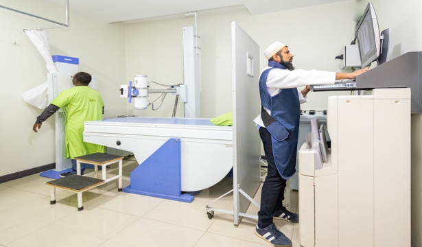
At Rayhaan Healthcare, we provide X-Ray services with the latest imaging technology, ensuring accuracy, safety, and fast results. Our hospital in Parklands, Nairobi, is equipped with cutting-edge radiology equipment to offer precise diagnostic imaging for a variety of medical conditions.
X-rays are an essential diagnostic tool used to view bones, tissues, and organs. Whether it’s for identifying fractures, infections, or other abnormalities, our highly skilled radiologists ensure that every scan provides clear, accurate images for proper diagnosis and treatment planning.
- Conditions Diagnosed with X-Rays:
- Bone fractures
- Joint dislocations
- Lung infections
- Digestive tract issues
- Dental conditions
- TEST OUTCOMES
- X-Ray Films
- Radiologist Report
- PATIENT PREPARATION
- Inform the radiographer if you are pregnant this is important information as it will make a difference in the way the X-ray is carried out or a different test altogether might be required. Your safety and that of your unborn child is our number one priority.
- A hospital gown will be given to you to wear, as some clothing can make it difficult to see the images clearly.
- You might also need to remove certain items, such as watches, necklaces and some types of clothing that contain metal objects, such as zips.
- If you are attending a follow-up X-ray to assess the progress of an injury or illness, you might need to carry any previous X-rays with you so that when reporting a comparison of the new X-ray with the old one can be done to see if there has been any recent changes.
Benefits of X-Ray Services at Rayhaan Healthcare:
We use modern X-Ray machines to provide high-resolution images, minimizing radiation exposure while ensuring quality results.
Our streamlined processes ensure that you receive your results promptly, allowing for timely diagnosis and treatment.
We provide a wide variety of payment options that includes mobile money, master card, & visa card.
We prioritize your comfort and safety throughout the procedure, ensuring a smooth and stress-free process.
At Rayhaan Healthcare, we are committed to delivering high-quality, personalized care. Our X-Ray services are designed to provide quick and reliable diagnostics to support your healthcare needs.
If you're in need of X-Ray services in Nairobi, visit Rayhaan Healthcare. We are conveniently located in Parklands, providing top-tier care to the local community. Call us on (+254)-111-051-530.